
PB 196
Description
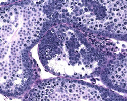
Disruption of spermatogenesis and accumulation of undifferentiated spermatogonia in GDNF overexpressing mice. Testicular morphology of GDNF-overexpressing mice is normal at birth. After 2 to 3 weeks, a chimeric histological pattern is observed. The tubular cross sections not only showed normal spermatogenesis but also displayed large cell clusters. Because these cells in the clusters did not show much nuclear heterochromatin and did express a spermatogonial marker, EE2, they could be morphologically classified as type A spermatogonia. The clusters gradually degenerated after puberty, resulting in tubular atrophy, and Sertoli cells phagocytosed the dead cells. At 10 weeks of age, only remnants of clusters were seen, but a rim of spermatogonia at the periphery of atrophic seminiferous tubules remained. No sperm was observed in seminiferous tubules or the epididymis.
MPATH / Pathology
MPATH 127 - atrophy
Gene
Gdnf
sex
Male
strain
FVB
organism
Mouse
EMAP / Embryonic stage, tissue or post-natal age:
99997 - Juvenile
genotype status
Homozygous
genetic manipulation
Transgenic-Ectopic-overexpression
MA / Anatomical Site
MA 412 - seminiferous tubule
Designated Allele Name
Experimental Manipulation
Further info
NOTE: Not all terms are currently shared between Pathbase and the above databases, some searches may not produce returns, in which case users should use synonyms or more inclusive text terms to search manually!
Copyright
This image remains the property of the originating Institution and should not be modified, reproduced or disseminated without the express permission of the submitter.
Gene Ontology
GO ID: 0005615 - extracellular space GO ID: 0009653 - morphogenesis GO ID: 0007399 - neurogenesis GO ID: 0007422 - peripheral nervous system development GO ID: 0030432 - peristalsis GO ID: 0007179 - TGFbeta receptor signaling pathway GO ID: 0005160 - transforming growth factor-beta receptor binding GO ID: 0007169 - transmembrane receptor protein tyrosine kinase signaling pathway GO ID: 0006916 - anti-apoptosis GO ID: 0007422 - peripheral nervous system development