-
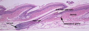 The tail skin in the adult mouse is very different from truncal skin. The epidermis is very thick and heavily cornified. The tail hair follicles are short and large compared to truncal hair follicles. A large, short hair fiber emerges and in the process uplifts the epidermis to create what looks like scales on the tail. In this saggital section there is a late anagen stage tail hair follicle in the center of the field. The new hair fiber pushes the old one (the club hair) to the side.
-
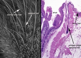 If one examines the hair around the anus of a mouse by scanning electron microscopy one will see a small cluster of very long and wide hair follicles in a cluster around the anus. These are in marked contrast to the very short truncal hairs that cover the rest of the body. Histologically, if one sections through the anus it is possible to find the very large hair follicles that are only located circumferentially around the anus. These follicles remain in anagen for long periods of time since the length of anagen is a direct relationship to the length of the hair fiber. The sebaceous glands are large compared to those in the normal truncal pilosebaceous units and are multilobulated. These may have a scent role as is the case in a number of other mammals.
-
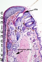 The eyelid protects the eye and also provides fluids for lubrication and barrier maintenance. The conjunctiva is a mucus membrane. The cilia is a specialized wide, short hair fiber produced by specialized hair follicles at the mucocutaneous junction. It is a pilosebaceous unit anatomically similar to truncal hair follicles. The large meibomian gland is located below this hair follicle and its duct emptied directly onto the conjunctiva.
-
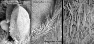
 Scanning electron microscopy of the mouse ear (pinna) demonstrates the relatively sparse, short hair on the inner and outer surface of its surface. Higher magnification illustrates the short, sparce nature of these hairs.
-
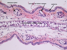
 The ear is a very specialized structure. It has a plate of auricular cartilage that runs down the center making histologic identification easy. The hair follicles found in this area are very short and thin with a very short anagen cycle. As such, usually hair follicles are found to be in prolonged telogen (resting phase), as is the case here. The epidermis is thin, although usually it is slightly thicker than that of the truncal skin.
-
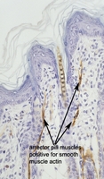 The skin from a young mouse has a thick epidermis as is seen here. There is a pigmented hair fiber emerging from one of the hair follicles. This section is labeled to demonstrate expression of the smooth muscle actin isoform. Note the long, thin, branching fibers near the epidermis. These are the arrector pili muscles. The elevate the hair fiber by pulling on the hair follicle. This happens during the "fight or flight" response in mammals when one's hair stands up. It is also important in thermoregulation. In the deeper dermis there are tubular structures that are also labeled. These are small arteries where their smooth muscle walls are labeled for this protein.
-
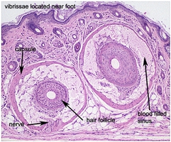 Vibrissae are specialized somatosensory organs in most mammals. Notable exceptions are humans that have no equivalent organ. While these are commonly seen on the muzzle and INCORRECTLY called whiskers (mice do not have whiskers) these can also be found near the feet. In this image there are two vibrissae cut in cross section. These are easily identified by the large blood filled sinus that surrounds the very large hair follicles.
Back to Top
 The tail skin in the adult mouse is very different from truncal skin. The epidermis is very thick and heavily cornified. The tail hair follicles are short and large compared to truncal hair follicles. A large, short hair fiber emerges and in the process uplifts the epidermis to create what looks like scales on the tail. In this saggital section there is a late anagen stage tail hair follicle in the center of the field. The new hair fiber pushes the old one (the club hair) to the side.
 If one examines the hair around the anus of a mouse by scanning electron microscopy one will see a small cluster of very long and wide hair follicles in a cluster around the anus. These are in marked contrast to the very short truncal hairs that cover the rest of the body. Histologically, if one sections through the anus it is possible to find the very large hair follicles that are only located circumferentially around the anus. These follicles remain in anagen for long periods of time since the length of anagen is a direct relationship to the length of the hair fiber. The sebaceous glands are large compared to those in the normal truncal pilosebaceous units and are multilobulated. These may have a scent role as is the case in a number of other mammals.
 The eyelid protects the eye and also provides fluids for lubrication and barrier maintenance. The conjunctiva is a mucus membrane. The cilia is a specialized wide, short hair fiber produced by specialized hair follicles at the mucocutaneous junction. It is a pilosebaceous unit anatomically similar to truncal hair follicles. The large meibomian gland is located below this hair follicle and its duct emptied directly onto the conjunctiva.
 Scanning electron microscopy of the mouse ear (pinna) demonstrates the relatively sparse, short hair on the inner and outer surface of its surface. Higher magnification illustrates the short, sparce nature of these hairs.
 The ear is a very specialized structure. It has a plate of auricular cartilage that runs down the center making histologic identification easy. The hair follicles found in this area are very short and thin with a very short anagen cycle. As such, usually hair follicles are found to be in prolonged telogen (resting phase), as is the case here. The epidermis is thin, although usually it is slightly thicker than that of the truncal skin.
 The skin from a young mouse has a thick epidermis as is seen here. There is a pigmented hair fiber emerging from one of the hair follicles. This section is labeled to demonstrate expression of the smooth muscle actin isoform. Note the long, thin, branching fibers near the epidermis. These are the arrector pili muscles. The elevate the hair fiber by pulling on the hair follicle. This happens during the "fight or flight" response in mammals when one's hair stands up. It is also important in thermoregulation. In the deeper dermis there are tubular structures that are also labeled. These are small arteries where their smooth muscle walls are labeled for this protein.
 Vibrissae are specialized somatosensory organs in most mammals. Notable exceptions are humans that have no equivalent organ. While these are commonly seen on the muzzle and INCORRECTLY called whiskers (mice do not have whiskers) these can also be found near the feet. In this image there are two vibrissae cut in cross section. These are easily identified by the large blood filled sinus that surrounds the very large hair follicles.

