Skinbase
Mouse Skin and Hair Database
Home
Pathbase
MGI
NARS
Nails
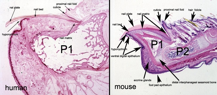
Saggital sections of human nails clearly reveals the marked similarities with mouse nails. Differences are more the angle and size (the human nail section is 10 times enlarged compared to the mouse). Human nails are dorsolaterally flattened to create a plate. Mouse nails are circumferentially layered or latterally flattened to produce more of a partial tube to protect the distal end of the phalanx. The major structures are found in both species. The nail matrix produces the nail plate which is attached to the phalanx by the nail bed. The nail plate normally separates from the nail bed where the hyponychium forms. This transition is characterized by the norcornified stratified squamous epithelium of the nail bed changing to the cornified (a stratum granulosum and stratum corneum form) hyponychium. The same anatomic structures are present in the mouse. It is therefore not surprising that changes in genes or damage to nails in mice result in exactly the same types of changes in human nails under similar circumstances. This is described in more detail and illustrated in the following book chapter: JP Sundberg and LE King. Skin and its appendages: normal anatomy and pathology of spontaneous, transgenic,and targeted mouse mutations. In: JM Ward, JF Mahler, RM Maronpot, and JP Sundberg (eds.) Pathology of Genetically Engineered Mice, Iowa State University Press, Ames, 2000, pp. 183-215.
Back to Top