Skinbase
Mouse Skin and Hair Database
Hair cycle
-
-
-
-
-
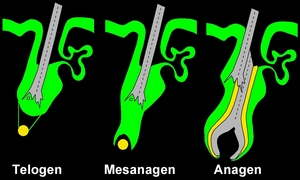
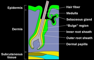
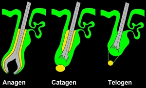
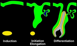
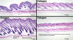 The new born mouse has a very thick epidermis and hair follicles that are still undergoing embryogenesis (top left panel). While little white fat is evident in the hypodermal fat region, oil red O stains can be helpful to identify adipocytes, which are still developing. Within 5 days the hair follicles produce fibers that emerge from the follicular ostium and are visible on the surface of the skin. At this stage and later (7 days of age, loweverleft panel) hair follicles are fully developed in late anagen. They are of different lengths because 4 different hair follicle types produce 4 distinct hair fiber types with different functions. The white adipose tissue is clearly evident and is usually markedly thicker at this stage than at any other as large amounts of energy are required to produce the hair fibers. After a defined period of time that varies between inbred strains and especially in many mutant strains or stocks, there is massive apoptosis and the bulbs degenerate and the remnants are pulled toward the epidermis. The basement membrane (commonly called the glassy membrane) becomes thick and pink in H&E stained sections. This will label in anagen with smooth muscle actin which is responsible for the contracture. The dermal papilla (sometimes called follicular papilla by those who do not study hair follicle biology) is retracted to the base of the sebaceous gland where the organ specific stem cells (bulge or wulst) is located. The resting telogen stage is short between the completion of embryogenesis and the start of the first adult hair cycle (classically and incorrectly called the second hair cycle). Length of telogen varies with age, usually getting progressively longer with successive hair cycles.
-
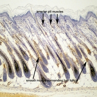 Skin sections labeled for expression of smooth muscle actin isoform reveal smooth muscle is present int he arrector pili muscles near the skin surface and surrounding the late anagen hair follicles in the basement (glassy) membrane as well as small arterial walls.
-
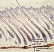 Immunohistochemical labeling of newborn mouse skin for the striated muscle actin isoform only labels the panniculus carnosus muscle below the fat layer and anagen stage hair follicles.
-
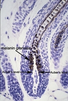 Pigmentation patterns are easily seen in hematoxylin stained skin sections of late anagen hair follicles in a C57BL/6J mouse. Note the abrupt line of where pigmentation starts. This is called Auber's level. Melanocytes inject melanin granules into keratinocytes. Because this only occurs in the anagen bulb in the mouse, it is easy to identify the anagen growth pattern by shaving mice and looking for the black patches (obviously only in pigmented mice).
-
 Higher magnification of the late anagen hair follicle illustrates the smooth muscle actin expression pattern around the outside of the follicle as well as the intenses expression around the small artery in the lower right of the field.

