-
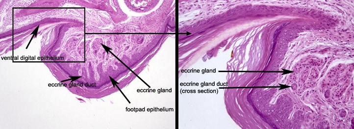
 The left panel is a low magnification of the bottom of the nail (ventral digital epithelium) and the foot pad which includes the eccrine gland. The duct is a coiled structure leading from the acini.
-
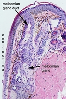 The mouse eyelid is a junction of the cornified stratified squamous epithelium of the haired skin and the noncornified stratified squamous epithelium of the conjunctiva, a mucus membrane. The meibomian gland is a large, modified sebaceous gland that produces lipids as part of the fluid barrier covering the cornea. The gland has a stratified squamous epithelial lined duct that opens through a slit-like opening onto the conjunctival surface.
-
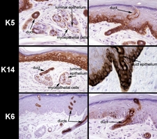 Mouse specific keratins 5 and 14 are expressed primarily in basal cells of the epidermis. This acid-base pair are also expressed in the eccrine gland duct epithelium and in flattened cells surrounding the acini (basal cells) that also are positive with actin markers indicating these are myoepithelial cells. Mouse specific keratin 6 is normally only expressed in the companion layer of the inner root sheath of the hair follicle. It is a nonspecific marker of epidermal hyperplasia in that it becomes expressed in the suprabasal epidermis when it is hyperplastic or neoplastic. In the eccrine glands this protein is expressed only in the luminal epithelium of the eccrine gland ducts.
-
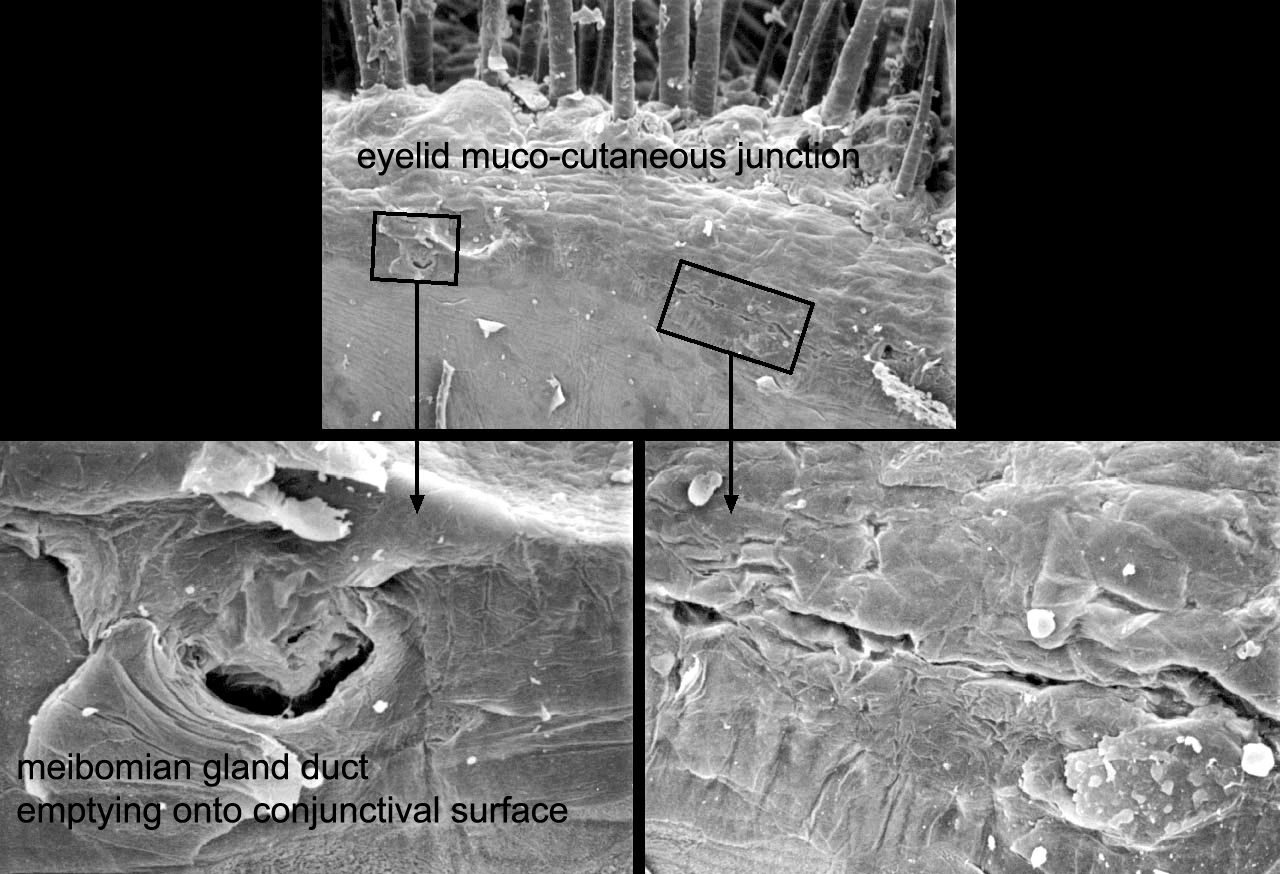
 The meibomian gland is a large, modified sebaceous gland that produces lipids as part of the liquid barrier protecting the conjunctiva. The ducts from these glands exit directly onto the conjunctiva. The ducts have slit-like openings near the mucocutaneous junction as indicated in the boxed areas. Higher magnification illustrates the features of these openings in these scanning electron micrographs.
RS Smith, JP Sundberg, SWM John. The anterior segment and ocular adnexae. In: RS Smith, SWM John, PM Nashina, and JP Sundberg. Systematic evaluation of the mouse eye. Anatomy, pathology, and biomethods. CRC Press, Boca Raton, FL, 2002, pp.3-23.
-
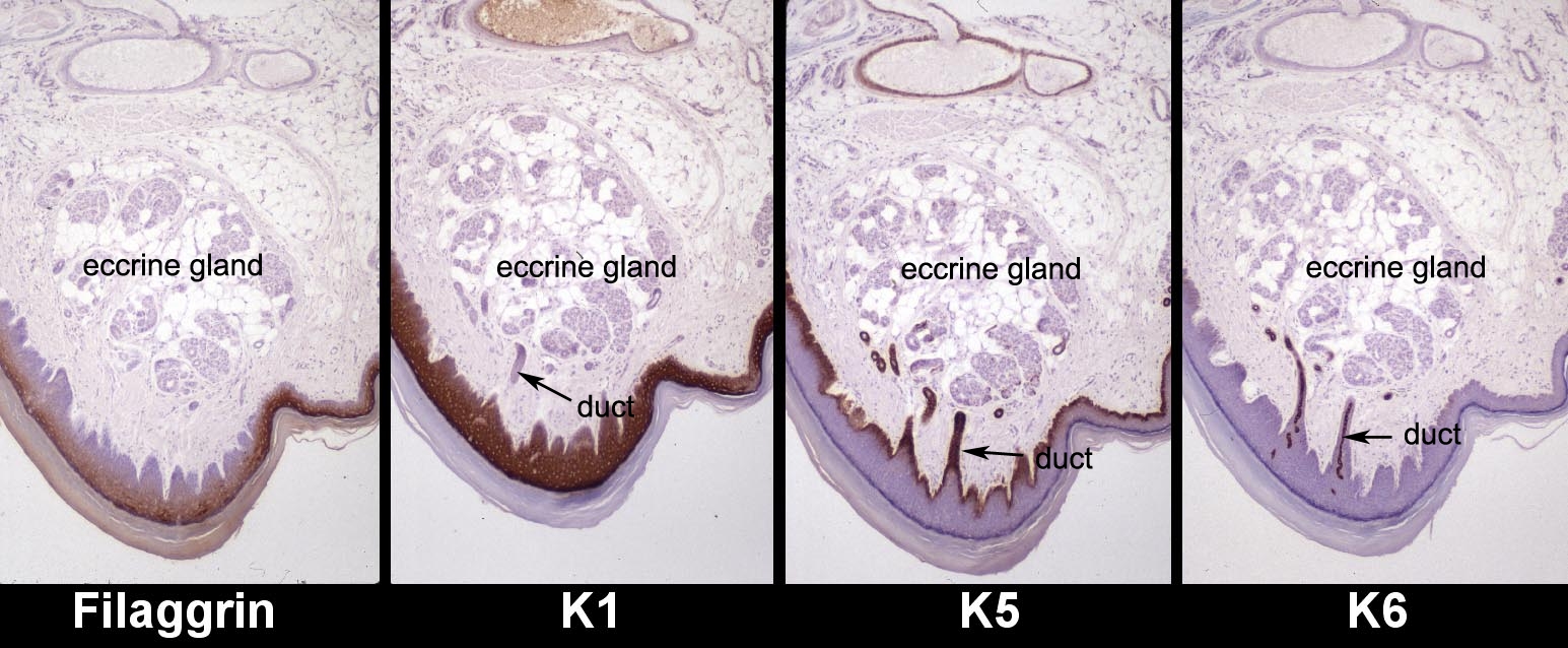
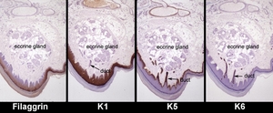 Serial sections of the mouse foot pad labeled with different mouse specific markers illustrate the variation in protein expression patterns. Filaggrin is the major protein found in the stratum granulosum of the epidermis. As seen in the left panel, this protein (brown) is only expressed in the upper layers of the foot pad epidermis. There is no filaggrin expression in the eccrine gland or duct. Another marker of terminally differentiated epidermal keratinocytes, keratin 1, is normally expressed in the suprabasal epidermal cells. This is the case in the second image. There is no expression in the eccrine glands or their ducts. By contrast, the two right panels illustrate keratins that are expressed in the adult eccrine glands and/or their ducts. Keratin 5 is normally expressed in the basal cells of the epidermis and mucus membranes. It is expressed in the basal cells of the foot pad epithelium as well as labeling the ducts and flattened cells surrounding the acini. The flattened cells are sometime called basal cells but are also called myoepithelial cells because they contain contractile elements (actin positive, not shown). Mouse specific keratin 6 is normally expressed in the companion layer of the inner root sheath of the hair follicle but not in the epidermis. It is a nonspecific marker of hyperplasia in mice and humans and squamous cell carcinoma (only mice) and under those circumstances it is expressed in the suprabasal epidermis. The the eccrine gland it is only expressed in the luminal cells of the eccrine gland ducts as seen in the far right panel.
-
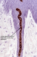 Keratin 6 is strongly and specifically expressed (brown) in the luminal cells of the eccrine gland duct. This protein is normally only expressed in the companion layer of the inner root sheath of the hair follicle.
Back to Top