

PB 24079
Description
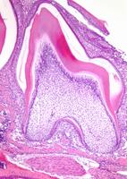
Partially erupted second molar. While crown is fully developed root formation is still ongoing. Due to decalcification during preparation the enamel is not seen, the space filled by enamel contains some non-degraded enamel matrix particularly seen in the distal portion near the cervix. The space defined by enamel is lined by reduced enamel epithelium, ie. ameloblasts. Outside this, stellate reticulum is present and limited by the outer enamel epithelium.
In the coronal portion, dentine formation is nearly complete and towards the pulpal cavity the mineralised dentine is lined by a thin layer of unmineralised pre-dentine. Bordering the pre-dentine, and slightly obliquely sectioned, odontoblasts are seen.
In the apical part the apices are still wide open and root formation is still ongoing. The apical part is delimited by Hertwig`s root sheath.
To the left in the image formation of alveolar bone is seen while in the space between the alveolar bone and the forming root is the developing periondotal ligament. At this stage the ligament is immature and densely populated by fibroblasts synthesising the collagen scaffold that provides connection between the root surface and the alveolar bone.
MPATH / Pathology
MPATH 458 - normal
Gene
sex
Male
strain
C57BL/6 CRL
organism
Mouse
EMAP / Embryonic stage, tissue or post-natal age:
99994 - Newborn
genotype status
Wild-type
genetic manipulation
None
MA / Anatomical Site
MA 1600 - lower jaw incisor
Designated Allele Name
Experimental Manipulation
Further info
NOTE: Not all terms are currently shared between Pathbase and the above databases, some searches may not produce returns, in which case users should use synonyms or more inclusive text terms to search manually!
Copyright
This image remains the property of the originating Institution and should not be modified, reproduced or disseminated without the express permission of the submitter.